Summary
Advanced imaging for TBI claims—such as 3T MRI, SWI, and DTI—offers clearer, more accurate diagnoses of hidden brain injuries. These tools help patients receive better treatment and give attorneys the evidence to win fair compensation. Learn how this technology strengthens both medical care and personal injury cases.
Table of Contents
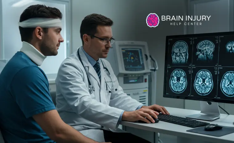
When you’ve suffered a traumatic brain injury in an accident, the invisible damage can be just as devastating as visible wounds. Traditional medical scans often miss subtle brain injuries that significantly impact your daily life and future earning capacity.
Fortunately, advanced imaging for TBI claims has revolutionized how doctors detect and document brain trauma. These cutting-edge technologies provide the detailed evidence to support your legal case and ensure you receive proper compensation for your injuries.
This blog will explain how advanced imaging works, why it matters, and how it helps patients, doctors, and legal teams.
Why Imaging Technology Matters in Brain Injury Cases
Traumatic brain injuries (TBIs) are often invisible on the surface. Traditional scans like CTs or standard 1.5T MRIs frequently miss subtle but severe internal damage. This is why courts, insurance companies, and medical experts turn to advanced imaging for TBI claims to detect, confirm, and support brain injury diagnoses.
According to the CDC, TBIs contribute to nearly 30% of all injury-related deaths in the U.S. Yet many survivors struggle to get proper care or compensation due to “normal” imaging results. That’s where 3T Magnetic Resonance Imaging, Diffusion Tensor Imaging (DTI), and Susceptibility Weighted Imaging (SWI) offer a deeper, clearer look at brain trauma with superior image quality and precise diagnosis capabilities.
The Game-Changing Difference: 3T vs. 1.5T MRI Technology
Not all MRIs are created equal. The “T” in MRI stands for Tesla, a unit of magnetic field strength. While most hospitals use 1.5T MRI machines, 3T MRI technology provides double the magnetic strength — resulting in sharper, more detailed imaging and crisper images that reveal subtle injuries traditional scans miss.
Feature | 1.5T MRI | 3T MRI |
Field Strength | 1.5 Tesla | 3 Tesla |
Image Quality | Moderate | High definition |
Scan Times | Longer | Shorter with faster scans |
Detection Ability | May miss microdamage | Better for subtle injuries |
The increased detail from this advanced MRI technology becomes crucial when working with the best brain injury attorney in Los Angeles. These clearer pictures provide compelling evidence that standard scans might miss entirely. The advanced scanner uses powerful magnetic waves and radio waves to create detailed imaging of soft tissues, blood vessels, and neural pathways without requiring contrast material in many cases.
As a noninvasive test, 3T MRI offers advantages over traditional imaging methods. The ultra-high fields generate superior signal clarity and utilize noise-reduction technology to produce accurate scans.
However, patients with large amounts of metal in their bodies may face restrictions, and some individuals may experience allergic reactions if contrast dye is necessary for enhanced visualization.
Susceptibility Weighted Imaging: Revealing Hidden Microbleeds
Susceptibility Weighted Imaging (SWI) represents a breakthrough in detecting microscopic brain hemorrhages. These tiny bleeds, invisible on conventional MRI, often occur during traumatic brain injuries when blood vessels stretch or tear during the initial trauma.
SWI technology works by detecting magnetic susceptibility differences between blood products and surrounding brain tissue. This specialized high-field procedure can identify:
- Microbleeds in white matter regions
- Small hemorrhages in gray matter areas
- Venous abnormalities following trauma
- Chronic blood deposits from previous injuries
For accident victims, SWI findings provide crucial documentation of brain trauma that might otherwise go undetected. This evidence becomes invaluable when pursuing compensation for cognitive difficulties, memory problems, or personality changes following your accident.
The detailed imaging capabilities of SWI create 3D pictures that clearly show vascular damage invisible to standard scanning methods.
Diffusion Tensor Imaging: Measuring Invisible White Matter Damage
Diffusion Tensor Imaging (DTI) measures how water molecules move through brain tissue. In healthy brains, water flows smoothly along white matter tracts. After trauma, this natural flow becomes disrupted, indicating microscopic damage to neural pathways that affects cognitive function and daily activities.
DTI technology reveals:
DTI Measurement | What It Shows | Legal Significance |
Fractional Anisotropy (FA) | White matter integrity | Cognitive impairment evidence |
Mean Diffusivity (MD) | Tissue organization | Memory loss documentation |
Axial Diffusivity (AD) | Axonal damage | Processing speed deficits |
Radial Diffusivity (RD) | Myelin disruption | Executive function problems |
This objective data helps medical experts explain why you experience concentration difficulties, memory lapses, or processing delays after your accident. DTI findings provide scientific backing for claims related to reduced work capacity or disability benefits, offering precise diagnosis capabilities that traditional imaging cannot match.
How These Imaging Tools Work Together
When used together, 3T MRI, SWI, and DTI provide a comprehensive, layered understanding of the brain’s structure and function:
- 3T MRI: Highlights physical abnormalities with high-resolution images and superior soft tissue contrast
- SWI: Detects microbleeds and small vessel injuries in blood vessels
- DTI: Reveals white matter damage that affects brain performance
Together, these scans offer patients a better diagnosis, allow doctors to create targeted treatment plans, and give attorneys the detailed evidence needed for fair compensation.
This trifecta of advanced imaging supports not just healing—but legal justice too. Unlike imaging for bone fractures or the musculoskeletal system, these brain-specific protocols focus exclusively on neural tissue damage and vascular injuries.
Benefits for Patients, Doctors, and Legal Teams
Advanced imaging isn’t just about stronger cases and better outcomes. Here’s what each group gains from this technology:
For Patients:
- More accurate diagnosis and treatment planning through clearer pictures
- Validation of cognitive and emotional symptoms
- Better understanding of recovery timelines
- Improved access to appropriate rehabilitation services
For Medical Teams:
- Enhanced ability to correlate symptoms with imaging findings
- Improved treatment protocol selection using detailed imaging
- Better patient communication about the extent of injury
- Stronger foundation for medical testimony with accurate scans
For Legal Representatives:
- Objective evidence supporting damage claims
- Scientific documentation of invisible injuries
- Enhanced ability to demonstrate future care needs
- Stronger position in settlement negotiations with precise diagnostic evidence
Statistical Impact on Legal Outcomes
Research shows that traumatic brain injury cases with advanced imaging documentation result in settlement amounts averaging 40-60% higher than those relying on standard scans alone. The detailed evidence these technologies provide helps establish the full extent of your injuries and their long-term impact on your life.
When to Request Advanced Imaging
If you’ve experienced any of these symptoms following an accident, discuss advanced imaging options with your medical team:
- Persistent headaches or dizziness
- Memory or concentration problems
- Personality or mood changes
- Sleep disturbances or fatigue
- Balance or coordination issues
- Sensitivity to light or noise
Don’t let insurance companies minimize your brain injury because standard scans appear “normal.” Advanced imaging for TBI claims can reveal the true extent of your trauma and support your right to fair compensation.
Taking Action After Your Brain Injury
The window for optimal brain injury documentation is limited. Advanced imaging becomes less definitive as time passes, making prompt action essential for your health and legal case.
Working with experienced legal counsel ensures you receive appropriate medical evaluations using the most advanced imaging technologies available. The right attorney will coordinate with medical experts who understand how to interpret these sophisticated scans and present findings effectively.
Make a Difference, Call Us Today
Advanced imaging technologies have transformed how we understand and document traumatic brain injuries. 3T MRI, SWI, and DTI provide the detailed evidence to validate your symptoms and support your legal claims. These tools reveal invisible damage that significantly impacts your life, even when traditional scans appear normal.
Don’t let inadequate medical documentation prevent you from receiving fair compensation for your brain injury. The sophisticated imaging for TBI claims available today can make the difference between a dismissed case and the financial recovery you deserve.
Ready to strengthen your brain injury claim with advanced imaging evidence? Contact Brain Injury Help Center today for a FREE CONSULTATION.
Our experienced legal team works with top medical professionals who utilize cutting-edge imaging technologies to document the full extent of your injuries. Call us now to protect your rights and secure the compensation you need for your recovery journey.